* So you had a dead cow and don't know why (CAUTION: pics!)
Jul 19, 2014 22:35:50 GMT -5
Claire, greatday, and 7 more like this
Post by milkmaid on Jul 19, 2014 22:35:50 GMT -5
*Mods: feel free to move to whatever appropriate place this belongs*
WARNING: Necropsy tutorial - graphic pictures below.
I've seen several posts by people perplexed why they had an animal die - especially the ones that appeared healthy the hour/day/week before. There's too many possibilities for any definitive diagnosis over the phone/internet/etc, especially with limited history and no pictures. Below is my attempt to help those type of posters narrow the possible differentials. Follow the instructions, take some good pictures, and you might be able to get an answer - or some good guesses - why your cow/calf/bull/etc died.
What do you need for a necropsy?
KNIFE - a sharp one! Most hunting knives and possibly some kitchen knives will work in a pinch. Mine is a 7" Jorgeson necropsy knife, available here: www.shopmedvet.com/product/necropsy-knife-narrow-blade-7inch The 5" blades will also work just fine, mine is a little excessive for calves.
GLOVES - please use!
CAMERA - pictures are essential
AX - you can cut through the ribs with a knife on anything up to about 18-24 months of age. Over that and you will need an ax (or massive set of bolt cutters/pruning shears) to get through the ribs.
TIME - I can do an 800lb calf alone in 7-12 minutes... but I did a couple a day for about 6 weeks to gain that competence. Plan on 45 minutes minimum with help, maybe an hour or two if alone or your first try at a necropsy. They're a little different than gutting a deer.
MISC - ruler if you need to describe something later, container(s) for specimens you're unsure about.
*Note: tissue samples go in the fridge, not freezer, and/or to your vet clinic ASAP*
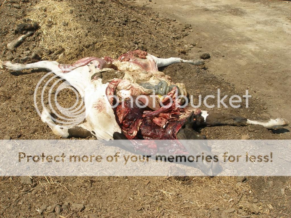
STEP 1: The dead animal can be positioned with its right side up, left side up, or on its back. Doesn't really matter. I prefer right side up as I was trained on a feedlot and since pneumonia is one of the top causes of death in cattle - it tends to affect the first lung lobe on the right before the others. If I open the calf with its right side visible, sometimes I have my answer as to the cause of death right away. The vet school's necropsy floor prefers left side up. I've traveled with a few veterinarians who preferred the calf on their back and splitting them down midline - my main complaint about that is it takes more than one person, a luxury I don't usually get.
STEP 2: Make a cut from the throat, down the neck, under the forelimb, and under the right hind limb. Lift the front leg and cut underneath it - there's just muscle and nerve attachments beneath it and it is fairly easy (dependent on size) to pick up and flip over. Depending on your size and strength - I've done some 1400 lb animals that I really did need an extra hand for leverage - or I've utilized chains, four wheelers, tractors, etc - use your head.

*Note: you can also make a cut along the hide up to the hip and skip cutting through the back leg as shown in the picture above - not quite as complete a necropsy and potentially more work later, but it is a valid option.
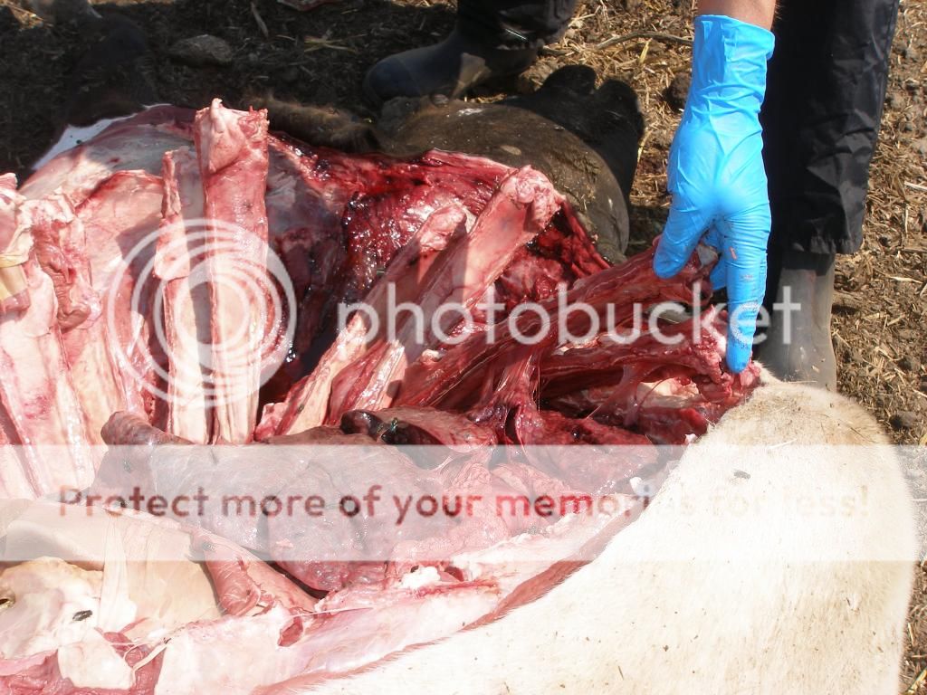
STEP 3: Getting through the ribs. Up to 18-24 months of age you can cut right through the costochondral junction with a knife.
I usually put a knife between each rib and separate them, then place my knife at the cartilage junction between ribs and sternum as shown in the pics above, and then just run my knife the length of the ribs. Younger animals its fairly simple, older animals can be incredibly frustrating if you don't get the knife set right. USE BOTH HANDS on the knife.
Here's your goal:

STEP 4: Look at the major organ systems and take pictures of all of them.
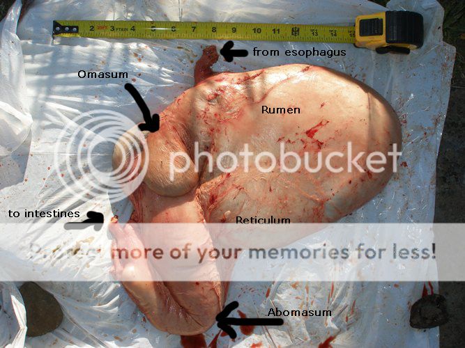
My minimum - lungs, heart, liver, kidney(s), rumen, omasum, reticulum, abomasum, esophagus, trachea, sometimes bladder, uterus, intestine, cecum, spleen depending on what I find up to that point/what the animal looked like prior to death. Always check the inside of the mouth for ulcerations (BVD can be the culprit).
*Note that in summer the mouth will often have white piles of fly eggs within a few hours after death - disgusting, but normal.
CUT ALL THE MAJOR ORGANS OPEN - don't just look at them. The interesting pathology is usually inside. This includes lungs, heart, liver, kidneys, esophagus, trachea as a bare minimum.
*Note: for the veterinarians on the board.... I do realize I'm describing a field necropsy and not a complete diagnostic laboratory one.
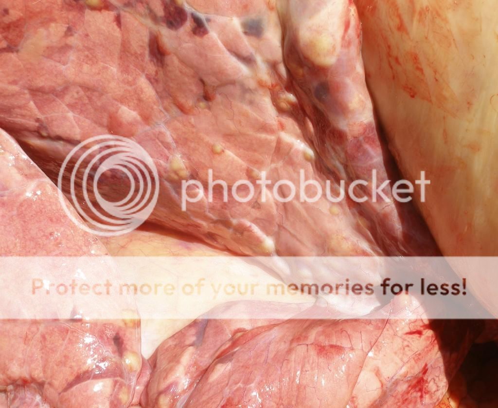
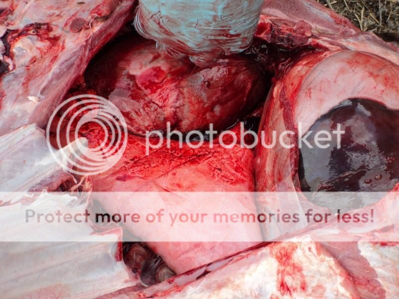
ABDOMEN: Evaluate the surface of the inside of the abdomen - this cow had peritonitis:

*Note that dragging a cow to a dead pile will occasionally cause some trauma that makes areas (on side close to ground) look bruised and sometimes look like peritonitis.
LUNGS: Look for adhesions between lungs and ribs as you break the ribs back.
Are they normal and pink or are there hard lobes, color changes, or abscesses in them?
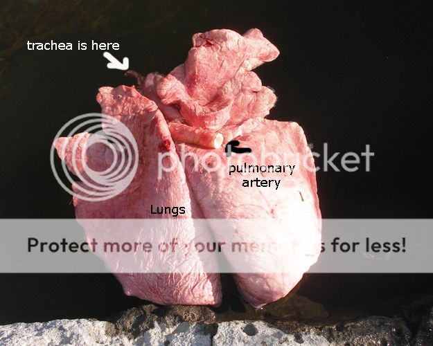
Split the trachea down to the terminal bronchioles - this animal had lungworms:

Look for fluid (clear, red, white) in the chest as you open the thorax to get to the heart and lungs. Sometimes you'll have a fountain of yellow fluid come spraying out when you slice through the ribs in a chronic pneumonia case.
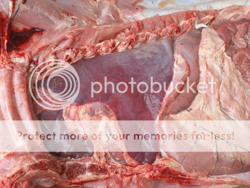
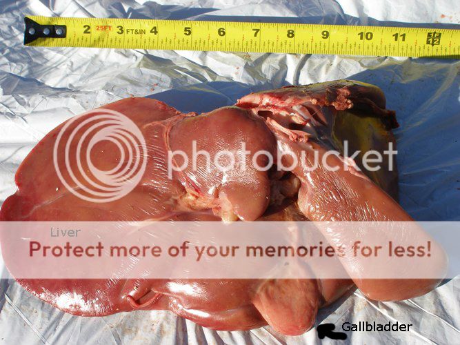
LIVER: Always cut slices (breadloaf style):
Look for spots that are inconsistent with the rest of the liver - this animal died of redwater following a necrotic area in the liver:

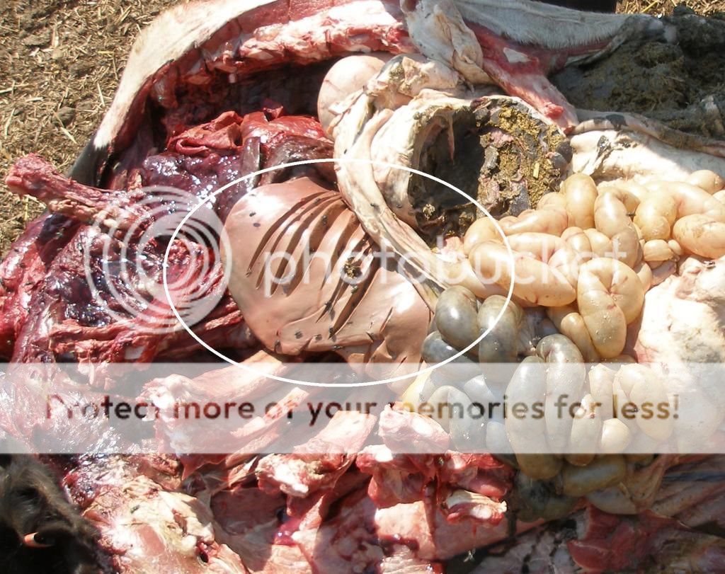
HEART: You can either cut it out with the lungs (remove the entire pluck with tongue, esophagus and trachea or just cut out heart and lungs together) or cut it out separately. This animal had a fascinating abscess attached to its heart (that's a normal looking bovine kidney next to it):
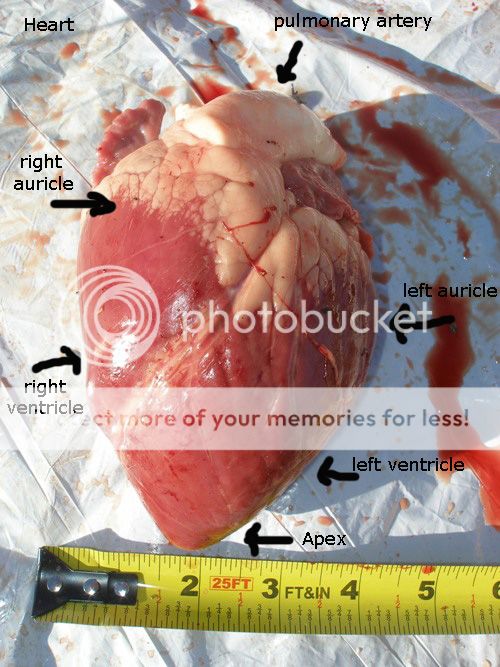
Always cut the heart open too. It has a right and left side - I usually split it top to bottom through each ventricle and up to the aorta and pulmonary arteries. Look for abscesses, color changes, surfaces that are not clean and shiny.
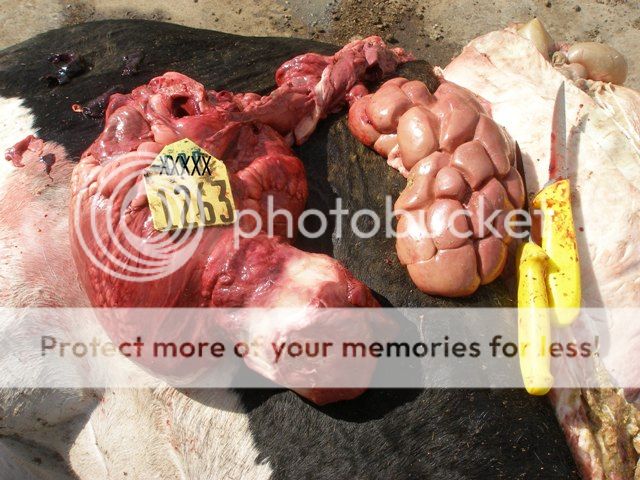
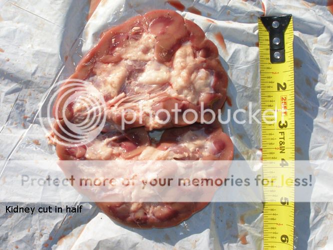
KIDNEYS: The left kidney is fairly easy to reach from this side and hides in the fat just behind the liver. It's lobed as can be seen in the picture above. Occasional problems with kidneys - 1) abscesses, 2) blocked (may be full of urine), or the interesting one below - she had a renal anurysm and bled out into the area surrounding the kidney.

ESOPHAGUS/TRACHEA: Some people take the tongue out as well, I'm not quite that motivated if I don't have a really good reason for it. I cut at the base of the tongue, peel the esophagus and trachea out together, slide my knife into the esophagus and split it (look VERY carefully for ulcerations not caused by your knife), then same for the trachea (BE CAREFUL - this is a good place to cut yourself). White foam is normal. I tend to make a short cut into the trachea, slide my knife between the rings to make a fingerhold, and then I can hold well enough to split the rest of the trachea.
Any ulcerations, surfaces that are not slick, thickened walls of the trachea, hemorrhage between tissues, etc are important and should be noted. Sometimes animals eat potatoes, onions, apples, or other food that can cause choking and death - be sure to check the length of the esophagus.
I'll get pictures at some point of this and add it to the post.
CALVES: Usually quicker and easier than adult animals, sometimes they have some really intriguing pathology.
LUNGS: If they're born dead, lungs tend to be dark pink/red and feel meaty. Born alive and they're pink and light. The ones below are normal.
This calf was born with a huge lung cyst - I don't recall the history well but believe it was born dead or died immediately after birth.

More normal ones on a 3 week old calf (note they are floating in water - good, normal lungs will float as pieces or in their entirety):
DOA - these lungs never inflated - if parts of lung on an adult animal look like this and feel thick/hard rather than light and squishy, think pneumonia.
DIGESTIVE TRACT:
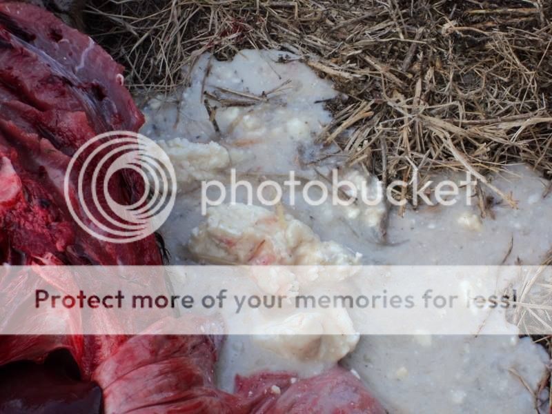
Normal for a 3 week calf:
Curdled milk in a milk-fed calf:
LIVER: Normal
HEART: Normal

KIDNEY: Normal

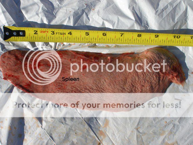
SPLEEN: Normal - you'll find this on the left side of the animal.
UTERUS: Normal for a 3 week old calf (open) - and it'll look very similar in any open heifer.

PICTURE TAKING TIPS:
#1: If you want answers, people need to be able to see context. Where is the item in the picture located in relation to other organs, how large is it, etc. It's better to take pictures a little too far away than too close.
#2: Either get the entire organ in the sun or get a shadow over it - it's difficult to tell actual color changes if half of it is covered by shadow.
#3: If it's covered in blood clots or dirt, it's incredibly hard to see whether its normal or not.
NECROPSY TIPS:
#1: Make sure you have a sharp knife.
#2: Cut them open ASAP after death - they'll decompose and organ systems won't look the same. 8 PM from death to 10 AM at necropsy is not ideal but dependent on cause of death, will usually still yield useful information. I did one of those just last week. 8 PM to 8 PM in the summer and you might be wasting your time. You might get 24 hours in the winter. Not in the summer.
#3: Cut EVERYTHING open. Feel free to post accompanying pictures of intact organs, but no lone pictures of an unscathed liver - breadloaf it so whatever is inside is visible. Same goes for lungs - cut a minimum of 3-4 slices into them - kidney, heart, etc. Some of my pictures are old and do not display this - just realize if you're posting pictures in search of answers it all needs to be cut if you want good responses.
#4: You'll miss more by not looking than by not knowing. Be curious and take more pictures than you think you'll need.
#5: If you have any concerns over diseases transmittable to humans - like anthrax in your area of the country - DO NOT open the animal.
#6: IF YOU PLAN TO HAVE DIAGNOSTIC TESTING DONE on the animal - e.g. you already know you have a disease outbreak, abortion storm, etc - let your veterinarian do the necropsy. Those types of testing may involve bacterial culture, something laboratories can't do when the same knife has been used to slice open the animal, digestive tract, affected organ(s), etc. This tutorial just covers curiosity.
QUESTIONS? - just ask. If you have something you know you're going to euthanize, feel free to ask here. If you're on the clock, your local vet clinic may be a much better option for immediate questions. Depending on your vet, most of the ones I've asked charge $75-100 to perform a necropsy and it may be helpful to watch one done once.
MM
WARNING: Necropsy tutorial - graphic pictures below.
I've seen several posts by people perplexed why they had an animal die - especially the ones that appeared healthy the hour/day/week before. There's too many possibilities for any definitive diagnosis over the phone/internet/etc, especially with limited history and no pictures. Below is my attempt to help those type of posters narrow the possible differentials. Follow the instructions, take some good pictures, and you might be able to get an answer - or some good guesses - why your cow/calf/bull/etc died.
What do you need for a necropsy?
KNIFE - a sharp one! Most hunting knives and possibly some kitchen knives will work in a pinch. Mine is a 7" Jorgeson necropsy knife, available here: www.shopmedvet.com/product/necropsy-knife-narrow-blade-7inch The 5" blades will also work just fine, mine is a little excessive for calves.
GLOVES - please use!
CAMERA - pictures are essential
AX - you can cut through the ribs with a knife on anything up to about 18-24 months of age. Over that and you will need an ax (or massive set of bolt cutters/pruning shears) to get through the ribs.
TIME - I can do an 800lb calf alone in 7-12 minutes... but I did a couple a day for about 6 weeks to gain that competence. Plan on 45 minutes minimum with help, maybe an hour or two if alone or your first try at a necropsy. They're a little different than gutting a deer.
MISC - ruler if you need to describe something later, container(s) for specimens you're unsure about.
*Note: tissue samples go in the fridge, not freezer, and/or to your vet clinic ASAP*
STEP 1: The dead animal can be positioned with its right side up, left side up, or on its back. Doesn't really matter. I prefer right side up as I was trained on a feedlot and since pneumonia is one of the top causes of death in cattle - it tends to affect the first lung lobe on the right before the others. If I open the calf with its right side visible, sometimes I have my answer as to the cause of death right away. The vet school's necropsy floor prefers left side up. I've traveled with a few veterinarians who preferred the calf on their back and splitting them down midline - my main complaint about that is it takes more than one person, a luxury I don't usually get.
STEP 2: Make a cut from the throat, down the neck, under the forelimb, and under the right hind limb. Lift the front leg and cut underneath it - there's just muscle and nerve attachments beneath it and it is fairly easy (dependent on size) to pick up and flip over. Depending on your size and strength - I've done some 1400 lb animals that I really did need an extra hand for leverage - or I've utilized chains, four wheelers, tractors, etc - use your head.
*Note: you can also make a cut along the hide up to the hip and skip cutting through the back leg as shown in the picture above - not quite as complete a necropsy and potentially more work later, but it is a valid option.
STEP 3: Getting through the ribs. Up to 18-24 months of age you can cut right through the costochondral junction with a knife.
I usually put a knife between each rib and separate them, then place my knife at the cartilage junction between ribs and sternum as shown in the pics above, and then just run my knife the length of the ribs. Younger animals its fairly simple, older animals can be incredibly frustrating if you don't get the knife set right. USE BOTH HANDS on the knife.
Here's your goal:
STEP 4: Look at the major organ systems and take pictures of all of them.
My minimum - lungs, heart, liver, kidney(s), rumen, omasum, reticulum, abomasum, esophagus, trachea, sometimes bladder, uterus, intestine, cecum, spleen depending on what I find up to that point/what the animal looked like prior to death. Always check the inside of the mouth for ulcerations (BVD can be the culprit).
*Note that in summer the mouth will often have white piles of fly eggs within a few hours after death - disgusting, but normal.
CUT ALL THE MAJOR ORGANS OPEN - don't just look at them. The interesting pathology is usually inside. This includes lungs, heart, liver, kidneys, esophagus, trachea as a bare minimum.
*Note: for the veterinarians on the board.... I do realize I'm describing a field necropsy and not a complete diagnostic laboratory one.
ABDOMEN: Evaluate the surface of the inside of the abdomen - this cow had peritonitis:
*Note that dragging a cow to a dead pile will occasionally cause some trauma that makes areas (on side close to ground) look bruised and sometimes look like peritonitis.
LUNGS: Look for adhesions between lungs and ribs as you break the ribs back.
Are they normal and pink or are there hard lobes, color changes, or abscesses in them?
Split the trachea down to the terminal bronchioles - this animal had lungworms:
Look for fluid (clear, red, white) in the chest as you open the thorax to get to the heart and lungs. Sometimes you'll have a fountain of yellow fluid come spraying out when you slice through the ribs in a chronic pneumonia case.
LIVER: Always cut slices (breadloaf style):
Look for spots that are inconsistent with the rest of the liver - this animal died of redwater following a necrotic area in the liver:
HEART: You can either cut it out with the lungs (remove the entire pluck with tongue, esophagus and trachea or just cut out heart and lungs together) or cut it out separately. This animal had a fascinating abscess attached to its heart (that's a normal looking bovine kidney next to it):
Always cut the heart open too. It has a right and left side - I usually split it top to bottom through each ventricle and up to the aorta and pulmonary arteries. Look for abscesses, color changes, surfaces that are not clean and shiny.
KIDNEYS: The left kidney is fairly easy to reach from this side and hides in the fat just behind the liver. It's lobed as can be seen in the picture above. Occasional problems with kidneys - 1) abscesses, 2) blocked (may be full of urine), or the interesting one below - she had a renal anurysm and bled out into the area surrounding the kidney.
ESOPHAGUS/TRACHEA: Some people take the tongue out as well, I'm not quite that motivated if I don't have a really good reason for it. I cut at the base of the tongue, peel the esophagus and trachea out together, slide my knife into the esophagus and split it (look VERY carefully for ulcerations not caused by your knife), then same for the trachea (BE CAREFUL - this is a good place to cut yourself). White foam is normal. I tend to make a short cut into the trachea, slide my knife between the rings to make a fingerhold, and then I can hold well enough to split the rest of the trachea.
Any ulcerations, surfaces that are not slick, thickened walls of the trachea, hemorrhage between tissues, etc are important and should be noted. Sometimes animals eat potatoes, onions, apples, or other food that can cause choking and death - be sure to check the length of the esophagus.
I'll get pictures at some point of this and add it to the post.
CALVES: Usually quicker and easier than adult animals, sometimes they have some really intriguing pathology.
LUNGS: If they're born dead, lungs tend to be dark pink/red and feel meaty. Born alive and they're pink and light. The ones below are normal.
This calf was born with a huge lung cyst - I don't recall the history well but believe it was born dead or died immediately after birth.
More normal ones on a 3 week old calf (note they are floating in water - good, normal lungs will float as pieces or in their entirety):
DOA - these lungs never inflated - if parts of lung on an adult animal look like this and feel thick/hard rather than light and squishy, think pneumonia.
DIGESTIVE TRACT:
Normal for a 3 week calf:
Curdled milk in a milk-fed calf:
LIVER: Normal
HEART: Normal
KIDNEY: Normal
SPLEEN: Normal - you'll find this on the left side of the animal.
UTERUS: Normal for a 3 week old calf (open) - and it'll look very similar in any open heifer.
PICTURE TAKING TIPS:
#1: If you want answers, people need to be able to see context. Where is the item in the picture located in relation to other organs, how large is it, etc. It's better to take pictures a little too far away than too close.
#2: Either get the entire organ in the sun or get a shadow over it - it's difficult to tell actual color changes if half of it is covered by shadow.
#3: If it's covered in blood clots or dirt, it's incredibly hard to see whether its normal or not.
NECROPSY TIPS:
#1: Make sure you have a sharp knife.
#2: Cut them open ASAP after death - they'll decompose and organ systems won't look the same. 8 PM from death to 10 AM at necropsy is not ideal but dependent on cause of death, will usually still yield useful information. I did one of those just last week. 8 PM to 8 PM in the summer and you might be wasting your time. You might get 24 hours in the winter. Not in the summer.
#3: Cut EVERYTHING open. Feel free to post accompanying pictures of intact organs, but no lone pictures of an unscathed liver - breadloaf it so whatever is inside is visible. Same goes for lungs - cut a minimum of 3-4 slices into them - kidney, heart, etc. Some of my pictures are old and do not display this - just realize if you're posting pictures in search of answers it all needs to be cut if you want good responses.
#4: You'll miss more by not looking than by not knowing. Be curious and take more pictures than you think you'll need.
#5: If you have any concerns over diseases transmittable to humans - like anthrax in your area of the country - DO NOT open the animal.
#6: IF YOU PLAN TO HAVE DIAGNOSTIC TESTING DONE on the animal - e.g. you already know you have a disease outbreak, abortion storm, etc - let your veterinarian do the necropsy. Those types of testing may involve bacterial culture, something laboratories can't do when the same knife has been used to slice open the animal, digestive tract, affected organ(s), etc. This tutorial just covers curiosity.
QUESTIONS? - just ask. If you have something you know you're going to euthanize, feel free to ask here. If you're on the clock, your local vet clinic may be a much better option for immediate questions. Depending on your vet, most of the ones I've asked charge $75-100 to perform a necropsy and it may be helpful to watch one done once.
MM